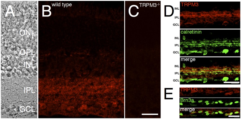
Fig 3. TRPM3 is expressed in the IPL and GCL of the mouse retina.

Mouse retina sections were labeled by immunofluorescence for TRPM3. A) Transmitted light DIC image showing the retinal cell layers. B) TRPM3 immunoreactivity is observed in the IPL and GCL of a WT mouse retina. C) No immunoreactivity is detected in a retinal section from a TRPM3-/- mouse. D) Transverse retinal sections of the inner retina double-labeled for TRPM3 (top, red) and calretinin (middle, green). Visible in the merged image (bottom), the outer, OFF half of the IPL (sublamina a) is more strongly labeled than the inner, ON, sublamina b. E) Imaging of the ganglion cell layer, double-labeled for TRPM3 (top, red) and Brn3a (middle, green). The merged image (bottom) shows that Brn3a-positive ganglion cells express TRPM3. Abbreviations are as follows: ONL, outer nuclear layer; OPL, outer plexiform layer; INL, inner nuclear layer; IPL, inner plexiform layer; GCL, ganglion cell layer. Scale bars = 20 μm.
