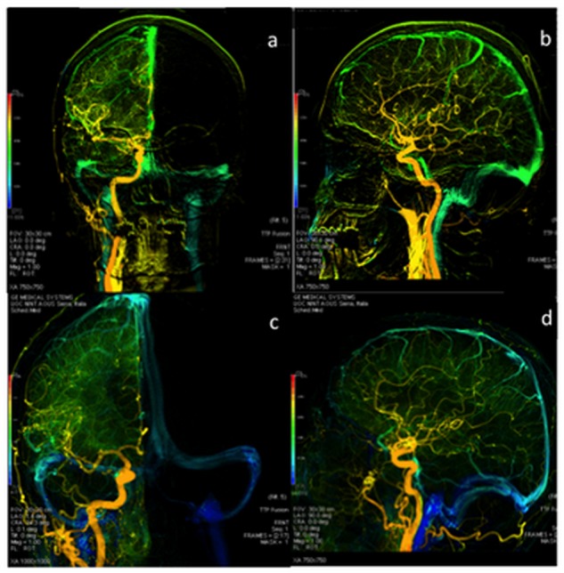
Fig 1. DSA examination: anterior-posterior and lateral views of color-coded right carotid artery of control (a;b) and MS patient (b;c).

The blue colour, shown only in MS patients, demonstrates a prolonged CCT. The veins are in green and not blue in the control.
