Figure 6.

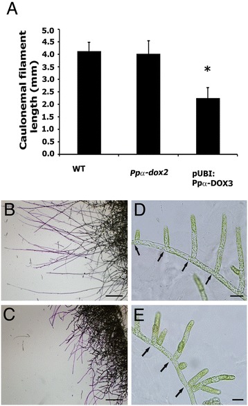
Effect of α-DOX-derived oxylipins on caulonemal development. (A) Values of average caulonemal filament length (in millimeters) measured in wild-type (WT), knockout line Ppα-dox-2 and overexpressing line pUBI:PpαDOX-3 colonies grown for 10 days in caulonemal induction conditions. Results and standard deviation corresponding to 16 colonies per sample are shown. (B) Border of a WT colony and (C) Border of a pUBI:Ppα-DOX-3 colony grown in caulonemal induction conditions showing toluidine blue stained caulonemal filaments. (D) Wild-type protonemal filaments at the border of a colony grown in BCDAT medium. (E) pUBI:PpαDOX-3 protonemal filaments at the border of a colony grown in BCDAT medium. Asterisk for caulonemal filament length from pUBI:Ppα-DOX-3 colonies, indicate that the values are significantly different from caulonemal filament length from WT plants according to Kruskal–Wallis test: P <0.001. Arrows in D and E indicate septa between cells. Scale bars represent 0,1 cm in B-C and 20 μm in D-E.
