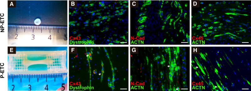
FIGURE 2.
Characterization of ETCs. SMBs and myotubes from nonpreconditioned engineered tissue construct (NP-ETC; A-D) and preconditioned engineered tissue construct (P-ETC; E-H) conditions. Macroscopic images of NP-ETC (A) and P-ETC (E). Immunohistochemical staining confirms increased gap junction (arrows, connexin43 [Cx43] and connexin45 [Cx45]) and adhesion protein (N-cadherin [N-Cad]) expression for P-ETC (F-H) compared with NP-ETC (B-D). The regression pattern of myotube-specific (dystrophin) and muscle-specific structural markers (α-actinin [ACTN]) was similar for both conditions. Scale bars = 50 μm.

