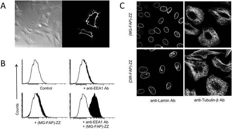
Figure 4.
FAP-ZZ affinity reagents target surface and intracellular proteins on fixed cells. A: Micrograph of fixed (non-permeabilized) HEK293 cells transiently expressing HAepitope at the surface. Cells were labeled with anti-HA antibody and (MG-FAP)-ZZ affinity reagent; then imaged with a 40× objective in presence of 50 nM MG-2p fluorogen. B: Flowcytometry histograms of fixed and permeabilized U937 cells labeled using anti-EEA1 (early-endosome-antigen-1) antibody and (MG-FAP)-ZZ affinity reagent. Each histogram represents a minimum of 10K live cell events in the presence of 25 nM MG-ester fluorogen. C:Micrographs of fixed and permeabilized HEK293 cells labeled with anti-lamin or anti-1-tubulin primary antibodies, and then by (MG-FAP)-ZZ or (DIR-FAP) ZZ secondary reagents. Images were acquired with a 60_ objective in the presence of 25 nM MG-ester or DIR fluorogens.

