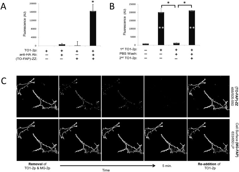
Figure 6.
Fluorescence signal manipulation from a FAP-ZZ affinity reagent. A: Fluorescence measurement graph of live HA-tagged U937 cells labeled with anti-HA antibody and (TO-FAP)-ZZ reagent in presence of 500 nM TO1-2p fluorogen. B: Graph of fluorescence measurement after addition, removal and re-addition of fluorogen in solution. Cells were labeled as in (A); then placed in fluorogen-free medium, and lastly, placed in medium containing 500 nM TO1-2p fluorogen (__corrected for background using unlabeled cells with fluorogen). C: Time-lapse micrographs of live HEK 293 cells HA-tagged to MG-FAP at the surface, and then labeled with anti-HA antibody and (TO-FAP)-ZZ affinity reagent. Cells were initially placed in medium containing both, 100 nM MG-2p and 500nM TO1-2p fluorogens; then placed in fluorogen-free medium, and lastly, placed in medium containing only 500 nM TO1-2p fluorogen.

