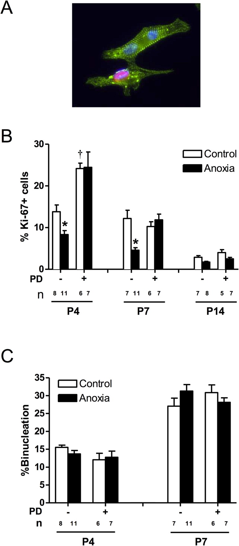
Fig 3. Effect of newborn anoxia and PD156707 on proliferation and binucleation of neonatal cardiomyocytes.
Cardiomyocytes were isolated from P4, P7, and P14 neonatal rats that were treated with control or anoxia, in the absence or presence of PD156707. Cells from P4 and P7 rats were stained with α-actinin and Ki-67, and nuclei were stained using Hoechst staining. P14 cardiomyocytes were stained with Ki-67 and analyzed via FACS. Panel A shows a representative image of cardiomyocytes stained with α-actinin (green), Ki-67 (red), and Hoescht (blue). Panel B shows percent of Ki-67 expressing cells. Panel C shows percent of binucleate cells. Data are means ± SEM. * P < 0.05, anoxia vs. control. † P < 0.05, -PD156707 vs. +PD156707. PD: PD156707; n: animal numbers.

