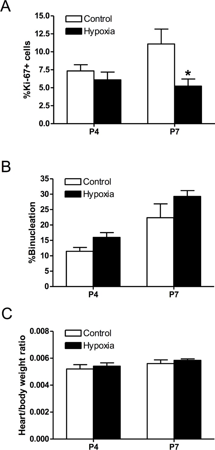
Fig 9. Effect of prenatal hypoxia on neonatal cardiomyocyte proliferation, binucleation, and heart to body weight ratio.
Cardiomyocytes were isolated from day 4 and 7 neonatal rats that were treated with control or maternal hypoxia. Cells were stained with α-actinin and Ki-67, and nuclei stained with Hoechst. Panel A shows percent of Ki-67 expressing cells (n = 3–4). Panel B shows percent of binucleate cells (n = 4). Panel C shows the heart to body weight ratio (n = 8–9). Data are means ± SEM. * P < 0.05, hypoxia vs. control.

