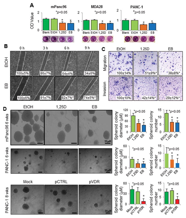
Figure 5.
Impact of altered VDR signaling on pancreatic cancer cell biology. Treatment with 1,25D and/or EB at 100 nM for 48 hours or as indicated inhibited the (A) growth of mPanc96, PANC-1, and MDA-28 cells in vitro (MTT assay); (B) horizontal migration of mPanc96 cells (gap-closing assay); (C) vertical migration and invasion of mPanc96 cells (Boyden chamber assay); and (D) stemness of PANC-1 and mPanc96 cells (spheroid colony formation assay). Moreover, transfection of PANC-1 cells with a VDR expression vector (pVDR) suppressed cell stemness more so than did transfection with a control vector (pCTRL). *P<0.01 as with the proper control.

