
Figure 1.
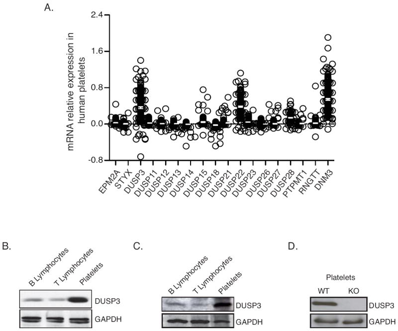
DUSP3 expression in human and mouse platelets. (A) Microarray data of mRNA expression of 17 atypical DSPs in human platelets isolated from 256 healthy volunteers. Each open circle represents one individual. DNM3 was used as positive control for platelet expressed mRNA. Data are presented as ratio of the fluorescence intensity for the DSP probe of interest and the mean fluorescence intensity for the housekeeping genes of each sample. A negative value corresponds to an expression bellow background level. Mean ± SEM are shown. (B-D) DUSP3 protein expression in human B and T lymphocytes and in platelets isolated from peripheral blood (B); in mouse splenic B and T cells and in washed platelets (C); and in WT and Dusp3-KO mouse washed platelets (D). Western blot analysis was performed using anti-human (B) and anti-mouse DUSP3 (C-D). GAPDH was used as loading control. Representative blots of three independent experiments are shown.