Figure 5.

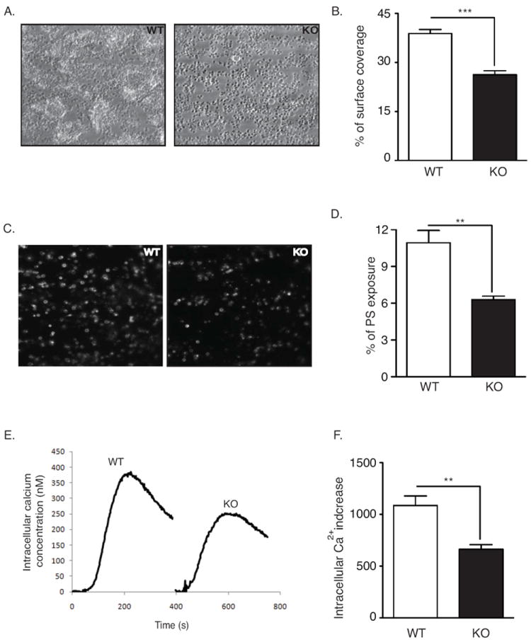
Impaired platelet aggregate formation on whole blood from DUSP3-deficient mice. (A-D) Anticoagulated blood from WT or Dusp3-KO mice was perfused over collagen-coated coverslip through a parallel-plate transparent flow chamber at a wall-shear rate of 1000 s-1 for 4 min. Representative phase-contrast images of fixed platelets (A) and percentages of surface coverage by platelets (B) are shown. Exposure of phosphatidylserine (PS) was detected by post-perfusion with heparin and OG488-labeled Annexin-V-containing rinsing buffer. Representative fluorescent images (C) and percentage of area coverage by labeled platelets (D) are shown. (E-F) Intracellular Ca2+ increase in WT and Dusp3-KO platelets upon CVX stimulation (50 ng/mL). Representative curves (E) and histogram depicting the area under the curve (F) are shown. WT values are arbitrarily set to 100%. Unpaired Student’s t-test was used for comparison. Results are representative of five independent experiments performed on platelet pools from three mice each. Data represent mean ± SEM of three independent experiments; **p<0.01, ***p<0.001.
