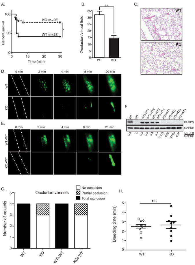
Figure 6.

Impaired arterial thrombosis and preserved hemostasis in DUSP3-deficient mice. (A-C) Pulmonary thromboembolism induced by injection of a mixture of collagen and epinephrine. Mortality incidence rates were compared using Kaplan-Meir with log-rank test (n=20 for KO and n=23 for WT mice) (A), quantification of the number of occluded vessels per visual field on lung sections from WT and Dusp3-KO mice (B), and representative field on lung sections from WT and Dusp3-KO mice (C) are shown. Data (from three to six lung sections from three mice of each group) were analyzed using unpaired Student’s t-test; **p<0.01. (D-E) FeCl3 injury of carotid arteries. Representative snapshot images in WT and Dusp3-KO mice (D), and in WT mice transplanted with WT (WT>WT) or with Dusp3-KO (KO>WT) BM cells (E) are shown. (F) Western blot analysis of DUSP3 expression in peritoneal TLs from WT>WT and KO>WT BM-transplanted mice used in the FeCL3 assay. Normalization was performed using GAPDH. (G) Numbers of intact and partially occluded vessels are shown for four mice of each group. (H) Tail bleeding time of WT (dark circle) and Dusp3-KO (open circle) mice. Each dot/circle represent one mouse. Results are presented as mean ± SEM.
