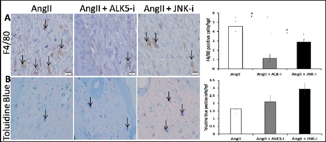
Figure 10. ALK5 inhibition decreases macrophage recruitment to the wound bed.
Representative 40× images (scale bars represent 50μm) of F4/80 (A) and toluidine blue (B) positive cells from wound samples of AngII only, AngII + ALK5 inhibitor, and AngII + JNK inhibitor treated mice on day 14 after wounding. Black arrows indicate representative positively stained cells. Quantitative analysis of F4/80 positive cells demonstrates a statistically significant lower density of macrophages in AngII + ALK5 inhibitor compared to other groups (*p<0.05). Quantitative analysis of Toluidine blue stained cells demonstrates no statistically significant difference between groups.

