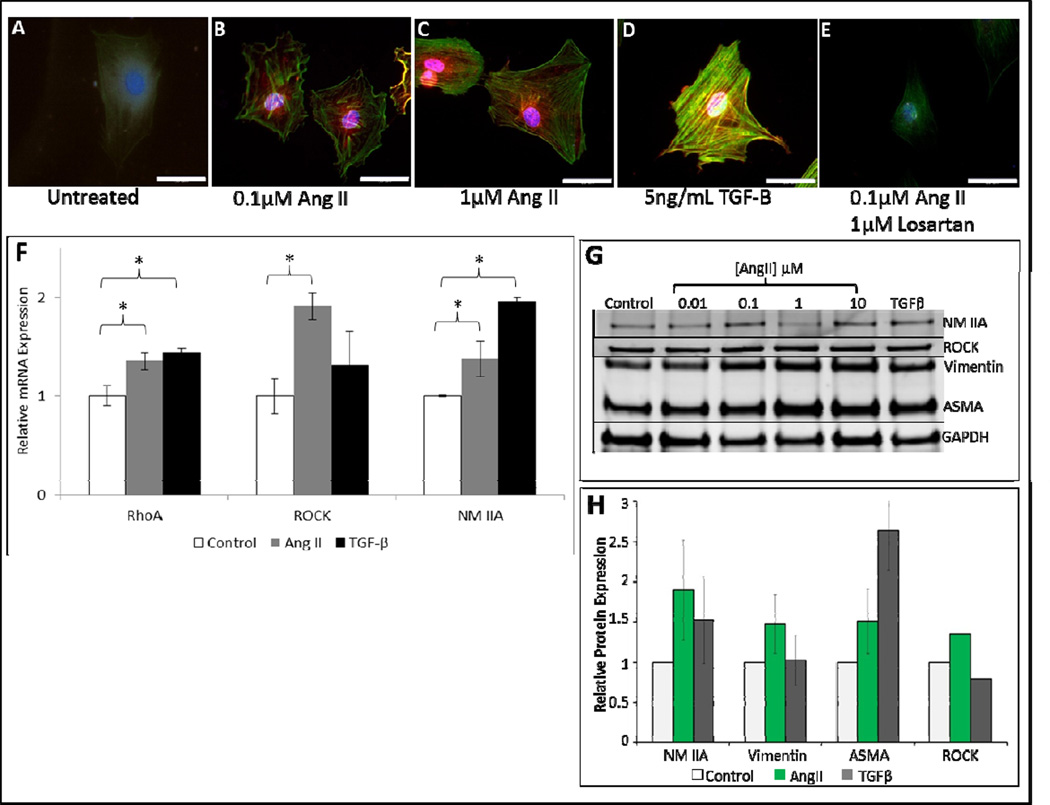
Figure 2. Angiotensin II upregulates expression of fibroblast contractile proteins.
Representative 200× images (scale bars represent 10μm), of filamentous actin phalloidin (FITC-phalloidin) and nuclei (DAPI) staining in 3T3s incubated in DMEM alone (untreated) (A), treated with 0.1μM AngII (B), 1.0μM AngII (C), positive control 5ng/ml TGFβ (D), and 0.1μM AngII + 1μM Losartan (E). Stress fiber formation is increased in AngII treated groups relative to groups incubated in DMEM alone (untreated). Losartan blocks AngII stimulation of stress fibers. qRT-PCR of control 3T3s (untreated), 3T3s treated with 0.1μM AngII, and 3T3s treated with 5ng/ml TGFβ as positive control (n=3) for 48 hours (F). Increased mRNA expression is seen in pro-contractile proteins RhoA, ROCK, and NM IIA of cells treated with AngII. Increased mRNA expression is seen RhoA and NM IIA of cells treated with TGFβ. Western blot analysis of NM IIA, Vimentin, αSMA, and ROCK in 3T3s treated with AngII (G). Quantitative analysis of western blot data for 0.1μM AngII treated cells demonstrating statistically significant increase expression of contractile proteins relative to control (H). Data shown is mean ±SEM, *p<0.05. Statistical significance was determined by ANOVA.

