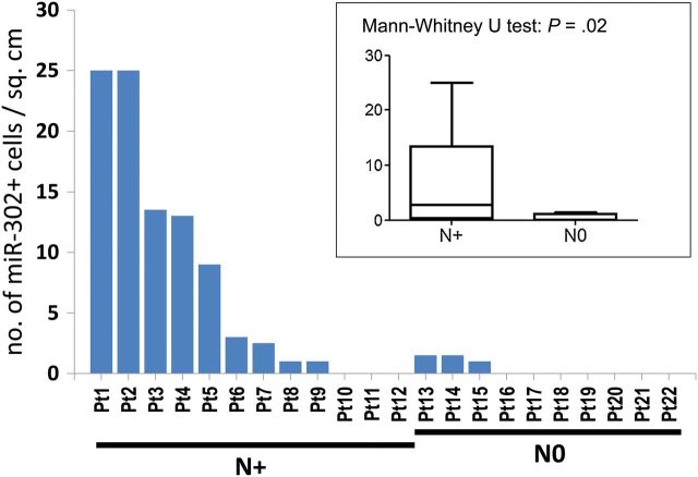
Figure 4.
MIR302 expression is higher in primary invasive ductal carcinoma with lymph node metastasis (test cohort). Primary tumors with lymph node metastasis contain larger amount of hsa-miR-302a positive cells, median = 2.75 positive cells/cm2, than those with no lymph node metastasis, median = 0 cells/cm2 (P = .02, two-tailed Mann-Whitney U test). Cancer tissues from 22 women with a history of invasive breast cancer were used. N+ on the abscissa indicate primary tumors with lymph node metastasis, N0 indicate those without metastasis. LNA in situ for hsa-miR-302a was performed on excisional biopsies. For multiple slides from the same biopsies the median scores were used. A patient who developed contralateral breast cancer was removed from the study. The insert box plot displays the number of hsa-miR-302a positive cells/cm2 for the lymph node positive and negative patients (error bars represent standard deviation).

