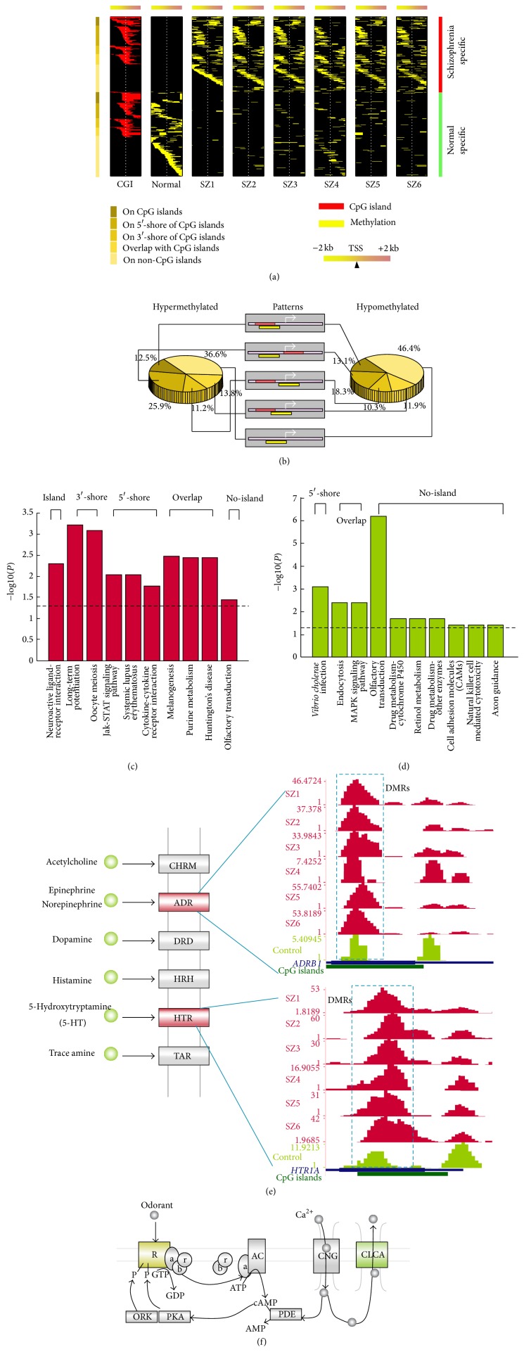
Figure 4.
Distinct DNA methylation patterns of promoters in schizophrenia (SZ). (a) Heat map of distinct patterns of promoter methylation. Each row represents a unique promoter region of 100 bp window size, covering ±2000 bp flanking the transcription start site, as indicated by the white dotted line. The location of a CGI (red) is shown in the first column. Promoters in the top panel are methylated in SZ while those in the lower panel are methylated in normal. Promoters are ordered by the location of methylation as represented with different shades of brown on the left. (b) Proportion of distinct patterns of promoter methylation. The middle panel shows a schematic diagram for distinct methylation patterns, while the left and right diagrams indicate the ratios of distinct methylation patterns for hyper- or hypomethylated promoters. (c) KEGG pathways are enriched by hypermethylated genes in SZ. The pathways are enriched by genes with distinct methylation patterns. (d) The KEGG pathways enriched in hypomethylated genes in SZ. (e) Neuroactive ligand-receptor interaction pathways enriched by two hypermethylated genes, Adrb1 and Htr1a. The subfigures show the methylation of these two genes in six SZ samples and one control. (f) The olfactory transduction pathway is aberrantly regulated in SZ; 3 hypermethylated genes are enriched in the receptor while 9 hypomethylated genes at the start and the end of this pathway are enriched.

