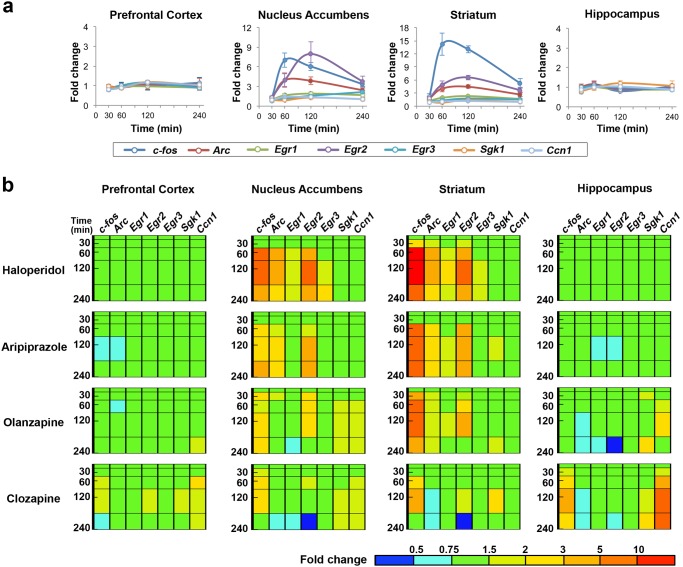
Fig 1. Antipsychotics induce distinctive IEG expression changes in nucleus accumbens and striatum, mainly due to their prominent D2R antagonistic activity.
(a) Time courses of seven IEG expressions induced by 0.3 mg/kg (p.o.) of haloperidol. Data are shown as mean± SEM (n = 5). Filled circles, p<0.05 in unpaired t-test with Welch’s correction, compound (n = 5) versus vehicle (n = 5) at the same time point; open circles, not significant. The changes of expression levels are shown as fold change over vehicle. (b) TFP of seven IEGs over time induced by antipsychotic agents in mouse brain. Antipsychotics; haloperidol (0.3 mg/kg, p.o.), aripiprazole (3 mg/kg, p.o.), olanzapine (10 mg/kg, p.o.) and clozapine (30 mg/kg, p.o.). The bar depicted in the bottom right corner represents the expression level of the gene searched in a TFP. Red represents high expression, green represents vehicle-control level expression, and blue represents low expression.

