
Fig. 1.
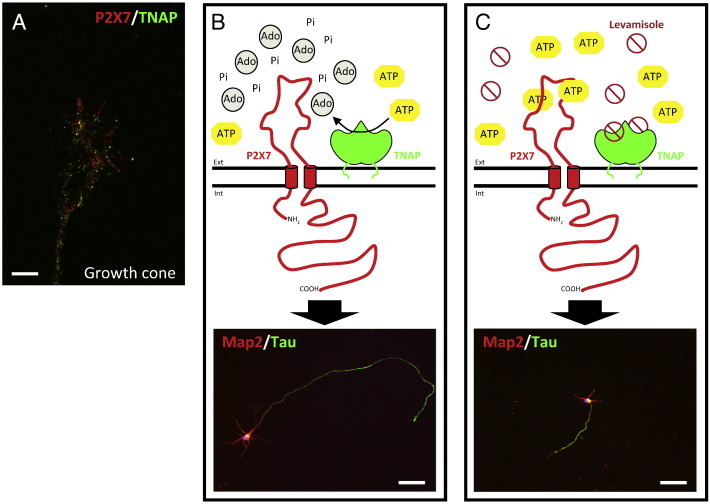
Schematic representation of the axonal growth regulation by the coordinated action of TNAP and P2X7R. A) Immunofluorescence image of the axonal growth of hippocampal neurons fixed at 3 DIV and stained with antibodies against TNAP (green) and P2X7R (red). The image shows the presence of both proteins at the growth cone of hippocampal neurons. Scale bar: 20 μm. B) TNAP hydrolyzes the physiological agonist of P2X7R, ATP, in the proximal environment of the receptor, which negatively regulates the activation of this receptor favoring in this way the axonal growth. C) The pharmacological inhibition of TNAP by levamisole produces an increase of ATP in the proximal environment of P2X7R, event that favor the activation of the receptor and then decreasing the axonal growth. The immunofluorescence images of lower panels show hippocampal neurons (3 DIV) stained with antibodies against axonal molecular markers, Map2 (red) and Tau (green), under normal condition (B) or treated with TNAP antagonist, levamisole, inhibiting axonal growth (C). Scale bar, B and C: 50 μm. Ext: extracellular space. Int: intracellular space. Ado: adenosine. Pi: inorganic phosphate. (For interpretation of the references to color in this figure legend, the reader is referred to the web version of this article.)