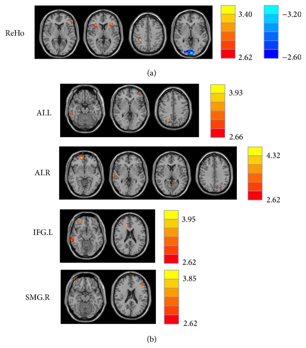
Figure 1.
(a) Regions of significant ReHo differences in tinnitus patients compared with healthy controls. Heat map (upper, right) shows areas of increased ReHo (t values 2.62 to 3.40; red to yellow, resp.) and decreased ReHo (t values −2.60 to −3.20; dark blue to light blue, resp.). Table 3 identified regions where significant increases and decreases occurred. (b) Significant increased functional connectivity of four seed regions between tinnitus patients and healthy subjects. Table 4 identified regions where significant increases occurred. The threshold was set at P < 0.01 (AlphaSim correction). L: left; R: right; ReHo: regional homogeneity; AI: anterior insula; IFG: inferior frontal gyrus; SMG: supramarginal gyrus.

