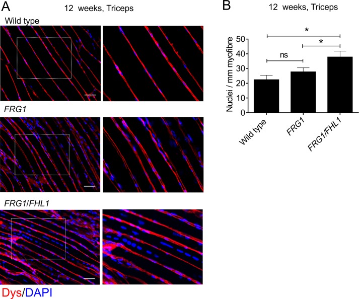
Fig 8. FHL1 enhances myoblast fusion in FRG1 mice.
(A) Representative images of longitudinal sections of triceps muscle from 12-week old wild type, FRG1 and FRG1/FHL1 mice co-stained for dystrophin to outline the muscle fiber membrane and DAPI to detect nuclei. Boxed region indicates area shown in high magnification image inset. Scale bars = 100μm. (B) The number of nuclei per mm of muscle fiber was counted as a measure of myoblast fusion at 12 weeks (wild type n = 3, FRG1 n = 4, FRG1/FHL1 n = 4). Data represent the mean ± SEM; ns not significant, *p<0.05determined by two-tailed Student’s T-test.

