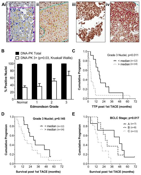
Figure 2. Increased DNA-PKcs protein levels in HCC and a shorter time to progression in patients receiving palliative TACE.
DNA-PKcs protein levels were determined immunohistochemically in 45 paired normal and HCC paraffin embedded tissues from cases described in Table 1. Hepatocyte nuclei were detected using an Aperio Imagescope nuclear algorithm. Level of nuclear expression was determined by pixel intensity, scored as un-elevated (0, blue), low (1, yellow), moderate (2, orange) or high (3, red). Application of the algorithm is shown in two paired normal and HCC tissues (a). DNA-PK was detected in the majority of hepatocyte nuclei and the % total nuclei positive (score 1-3) is shown, as % of nuclei scoring 3+ within each Edmondson tumour grade (normal n=45, grade 1 n=13, grade 2 n=23, grade 3 n=9). Grade 3+ nuclei increased significantly with tumour grade (b). Time to radiological progression (TTP) in patients receiving first line treatment with doxorubicin TACE (patients without extra-hepatic disease, no portal vein thrombosis, Childs-Pugh grade A, unsuitable for 1st line RFA) is shown in (c), where ‘High DNA-PKcs’ cases were those with > 48% HCC nuclei grade 3+ (n= 14; range 48-99%), compared to ‘Low DNA-PKcs’ cases, with <48% grade 3+ (n=12; range 0-43%). Median TTP was 4.5 versus 16.9 months (p=0.011 Kaplan Meier). Survival within this selected group was not significantly associated with any individual factor, including nuclear DNA-PK level (d), but was predicted by BCLC stage A-C, combining tumour, liver function and performance status (P=0.017 Kaplan Meier) (e).

