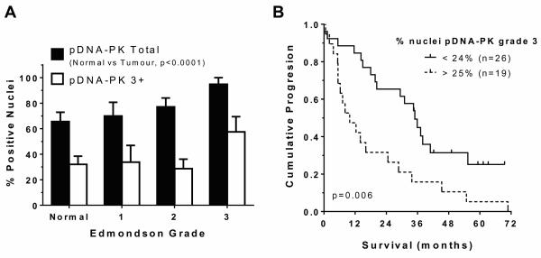
Figure 3. Increased tumour pDNA-PK and poorer survival.
Immunohistochemical pDNA-PKcsserine2056 levels were determined by pixel intensity, scored as described in Figure 2. Compared to total DNA-PK, fewer nuclei scored positive for pDNA-PKcsserine2056 overall. The % mean±SEM of positive total nuclei (scores 1-3 combined) and nuclei scoring 3+ within each Edmondson tumour stage are shown in (a). Panel (b) shows median survival of patients grouped as high (>25% 3+ nuclei, n=19) was 9.9 months, compared to low (<25% 3+ nuclei, n=26) tumour pDNA-PK (35 months; p=0.007, Kaplan Meier, p=0.006 by multivariate Cox Regression, including other tumour factors that were significant by univariate analysis: tumour size, tumour number, presence of extra-hepatic disease, presence of portal vein thrombosis).

