Figure 5.
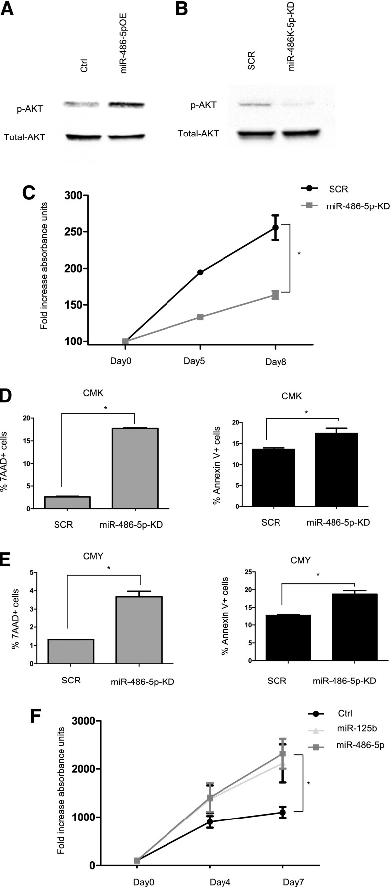
mir-486-5p enhances survival of ML-DS cells. (A-B) Western blot analysis of p-AKT levels (A) in CMS cells overexpressing miR-486-5p compared with empty vector (Ctrl) (B) in CMK cells after miR-486-5p KD compared with SCR. (C) MTT assay in CMK cells after miR-486-5p KD compared with SCR. *P < .01 (2-tailed Student t test). (D-E) FACS analysis of 7AAD and Annexin V positive cells in CMK (D) and CMY cells (E) after miR-486-5p KD compared with SCR. (F) MTT assay in CMS cells overexpressing miR-486-5p and miR-125b (positive control for growth-promoting miR18) or empty vector (Ctrl). *P < .05 (2-tailed Student t test) for each miR. SCR, scrambled.

