
Fig. 2.
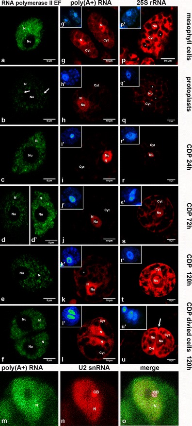
a–f Localisation of RNA polymerase II elongation form, g–l poly(A+) RNA, p–u 25S rRNA transcripts and m–o co-localisation of poly(A+) RNA with U2 snRNA in leaf mesophyll cells, protoplasts and CDP of Arabidopsis thaliana. a Nucleus isolated from leaf mesophyll cells. A homogenous fluorescence signal occurs in the entire nucleoplasm, and the nucleolus is devoid of signal. b Nucleus from a protoplast immediately after isolation (0 h). A weak fluorescence signal is visible in the form of different sizes of foci (indicated by an arrow). c Cell nucleus at 24 h after isolation. RNA POL II EF foci occur in the entire nucleoplasm. d CDP after 72 h showing a cell with weak staining similar to protoplasts. d′ Cell nucleus 72 h after isolation and right before cell division. The distribution of RNA POL II EF is comparable with that observed in nuclei isolated from leaf mesophyll cells. e Nucleus from a non-divided cell at 120 h after isolation. Easily distinguishable RNA POL II EF foci can be observed. f Two daughter nuclei after cell division show RNA POL II EF distributed homogenously. The fluorescence is weaker than in the 72-h CDP. g–l Localisation of poly(A+) RNA transcripts by FISH. g Leaf mesophyll cells show strong, homogenous labelling of the nucleoplasm; a weaker signal is observed between chloroplasts in the cytoplasm. h Protoplast with poly(A+) RNA distribution similar to mesophyll cells. i CDP cultured for 24 h. Staining is increased in the nucleus and decreased in the cytoplasm compared to a protoplast. A few aggregates are visible in the cytoplasm, while most of the signal is concentrated in the nucleus. j CDP after 72 h have a homogenous signal that is weaker than in the 24-h cultured CDP and is observed mainly in the cell nucleus. k A CDP cultured for 120 h is not yet divided and shows a strong homogenous signal in the nucleus; in the cytoplasm, the signal is diffused between cell compartments. l Divided cells after 120 h of culture show strong labelling in the nucleus. A newly synthesised cell wall is visible between cells. g′–l′ DAPI staining (blue) of cells in g–l. m–o Co-localisation of poly(A+) RNA and U2 snRNA (red) in nuclei of CDP cultured for 120 h. m Poly(A+) RNA signal occurs both in the nucleoplasm and cytoplasm. n The U2 snRNA signal is present mainly in the nucleus. In the nucleus, U2 snRNA is distributed homogenously in the nucleoplasm with signal aggregation representing a Cajal body (CB). o A merge shows no co-localisation of poly(A+) RNA and U2 snRNA in CBs. p–u Localisation of 25S rRNA transcripts by FISH. p Cell from leaf mesophyll tissue with fragments of adjacent cells. A strong, homogenous signal is observed in the nucleolus and in the spaces between chloroplasts. q Protoplast with decreased fluorescence intensity in the entire cell compared to a mesophyll cell with no changes in the distribution pattern. r A very weak fluorescence is observed in the cytoplasm in CDP after 24 h of cell wall removal. s CDP cultured for 72 h shows visible large nucleolus in the centre of the cell. The signal re-appears in the cytoplasm and occurs in spaces between plastids and under the cell membrane. t Non-divided CDP after 120 h of culture. A large, strongly labelled nucleolus is visible in the centre of the cell. A very intense and homogenous signal is evident in the cytoplasm. u Two divided cells after 120 h of culture. A visible, strong, homogenous signal in the cytoplasm is especially concentrated near the new cell wall (indicated by an arrow), and the signal is diffused in nucleoli and dispersed in the nucleoplasm. p′–u′ DAPI staining (blue) of cells in p–u. N nucleoplasm, Nu nucleolus, Cyt cytoplasm, * chloroplasts/plastids