Figure 2.
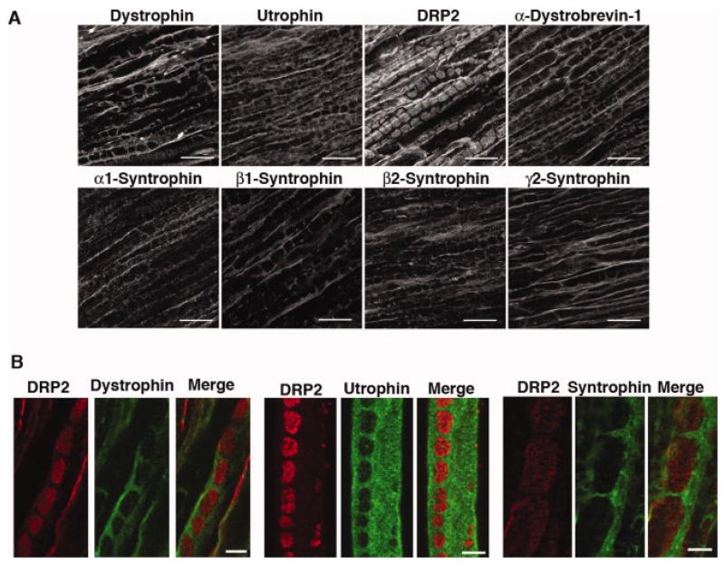
Localization of DGC proteins to Cajal bands and plaques and comparison of the distribution of DRP2 with dystrophin, utrophin, and α/β syntrophin in sciatic nerve. (A) Longitudinal sections of mouse sciatic nerve were stained with the indicated antibodies. Only DRP2 is localized to plaques. Dystrophin, utrophin, α-dystrobrevin-1, and α-, β1-, β2-, and γ2-syntrophin are all restricted to Cajal bands. Scale bar is 10 μm. (B) DRP2 (red) is localized to plaques while dystrophin, utrophin and α/β syntrophin (visualized with mAb 1351) (green) are restricted to Cajal bands. Right and left panels, rat sciatic nerve longitudinal sections. Middle panel, teased fiber. Scale bar is 5 μm.

