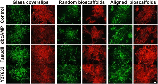
Figure 1.
Effect of drug treatments on morphology and expression of GFAP and AHNAK in astrocytes cultured on different substrates. Astrocytes were treated for 72 h with vehicle (Control), dbcAMP (1 mM), Fasudil (100 μM) or Y27632 (30 μM) on glass coverslips, random or aligned bioscaffolds and immunostained to reveal GFAP (green) or AHNAK (red). Paired images represent the same field. Scale bar = 50 μm.

