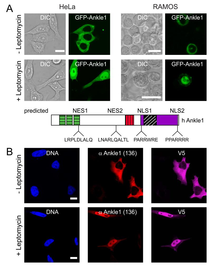
Fig. 4. Human Ankle1 shuttles between nucleus and cytoplasm.
(A) GFP-tagged Ankle1 was expressed in HeLa or RAMOS cells and imaged by live cell microscopy (GFP) and differential interference contrast (DIC) microscopy before (-) and after (+) treatment with Leptomycin for one hour. Nuclear export and nuclear localization signals identified by in silico prediction software are indicated in the cartoon (NES, NLS). (B) HeLa cells were transfected with hAnkle1-V5, and processed for immunofluorescence microscopy before and after three hours of Leptomycin treatment. Bars, 10 μm.

