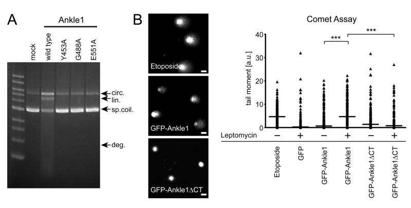
Fig. 5. Ankle1 cleaves DNA in vitro and in vivo.
(A) Purified wildtype and mutated HAStrep-Ankle1 expressed in HEK cells was incubated with supercoiled pFastBac1 plasmid and DNA species were separated by agarose gel electrophoresis. Arrows indicate different intermediate steps of plasmid degradation (o.circ=open circle, lin=linear/nicked, sp.coil=supercoiled, deg=degraded). (B) Representative images of ethidiumbromide stained nuclei in a Comet assay. Bars indicate 10 μm. Comets were measured using the Comet Assay IV software (Perceptive Instruments, Haverhill, UK), quantified and visualized in Graphpad Prism. Median values are indicated as horizontal lines. *** indicate statistically significant differences in median values between populations (p<0.001). Comet formation (i.e. DNA fragmentation) was evaluated in single cells of three independent cell populations (n>50 each).

