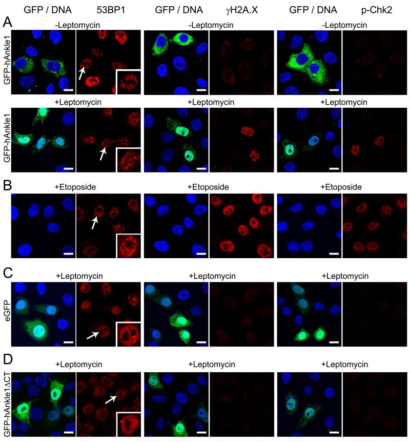
Fig. 6. Accumulation of ectopic Ankle1 in the nucleus causes DNA damage.
HeLa cells were transiently transfected with GFP-Ankle1 (A), GFP (C) or GFP-Ankle1ΔCT (D) or incubated with Etoposide (B), and treated with Leptomycin as indicated. All samples were processed in parallel for confocal immunofluorescence microscopy. Fixed cells were stained for 53BP1, γH2A.X or phosphorylated Chk2 using specific antibodies (all red), DNA was stained with DAPI (blue), GFP and GFP-fusion proteins are shown in green. Bars indicate 10 μm; arrows, cells shown in the insets. Representative images out of >3 independent experiments.

