
FIGURE 1.
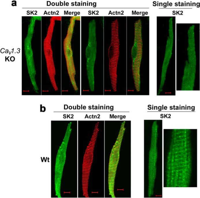
Subcellular localization of SK2 channels was altered in atrial myocytes isolated from homozygous Cav1.3 null mutant mice. The subcellular distribution of SK2 channels in atrial myocytes isolated from Cav1.3−/− mice (KO) (a) as compared with those of the WT animals (b). Anti-SK2 antibodies were used for single staining. The double staining showed SK2 channels and α-actinin2 (Actn2) localization patterns. The right panels in a and b show single staining at higher magnification. Scale bars are 10 μm. Atrial myocytes were obtained from three pairs of Cav1.3−/− mice compared with WT littermates. All experiments were repeated independently three times and consistent data were obtained as shown.