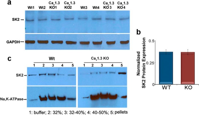
FIGURE 2.
Subcellular distribution of SK2 channel proteins in atrial tissues from Cav1.3−/− (KO) and WT mice. a, Western blot analyses showing similar expression levels of SK2 channel protein in whole atrial tissue lysates isolated from Cav1.3−/− and WT animals. GAPDH was used as a loading control. b, summary data for SK2 protein expression levels normalized to GAPDH. c, abnormal distribution of SK2 channels was observed in Cav1.3−/− mice using a discontinuous sucrose density gradient ultracentrifugation. The purity of membrane fractionation in mouse atrial tissues was tested and normalized by Na,K-ATPase as the plasma membrane marker. Each sample of atrial myocytes was isolated from 5 animals and the experiments were repeated independently three times.

