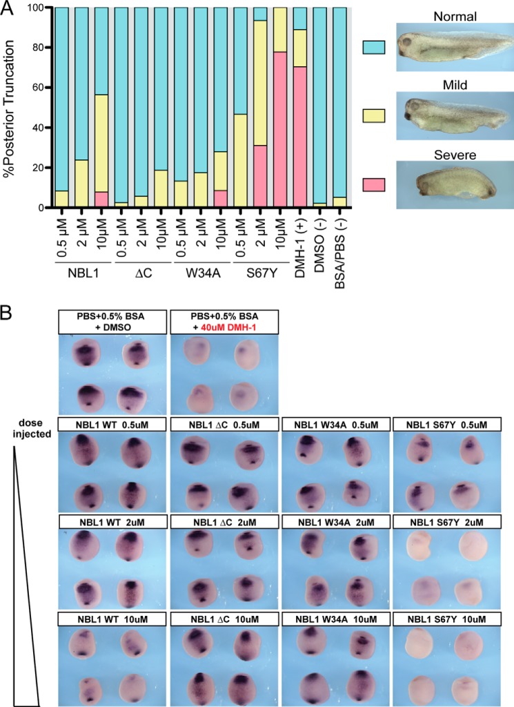
FIGURE 7.
Analysis of NBL1 and NBL1 mutants in vivo. In vivo BMP inhibition activity of NBL1 and various mutants tested during Xenopus embryo development. The purified proteins (0.5, 2, and 10 μm) were injected into the blastocoel cavity of stage 9 embryos. A, bar graph represents embryos that were scored for defects in BMP-dependent axial development at stage 35 using the standard dorsoanterior index and classified into different subgroups based upon the severity of posterior truncation. Each bar represents 100% percent of the embryos tested where cyan represents the percent of the population with a normal phenotype, yellow represents a mild phenotype (mild tail truncation), and red represents a severe axial truncation. Images show representative examples of each described phenotype. B, embryos at stage 20 (ventral views) were evaluated by in situ hybridization for mRNA expression of the direct BMP-target gene sizzled. In control embryos sizzled expression is detected in the ventral mesoderm as a result of endogenous BMP signaling (BSA/PBS control), whereas strong inhibition of endogenous BMP signaling results in very little to no sizzled expression (DMH-1 control). Each protein was tested in at least 3 separate injection experiments, with 80–100 embryos tested in total for each.

