FIGURE 6.
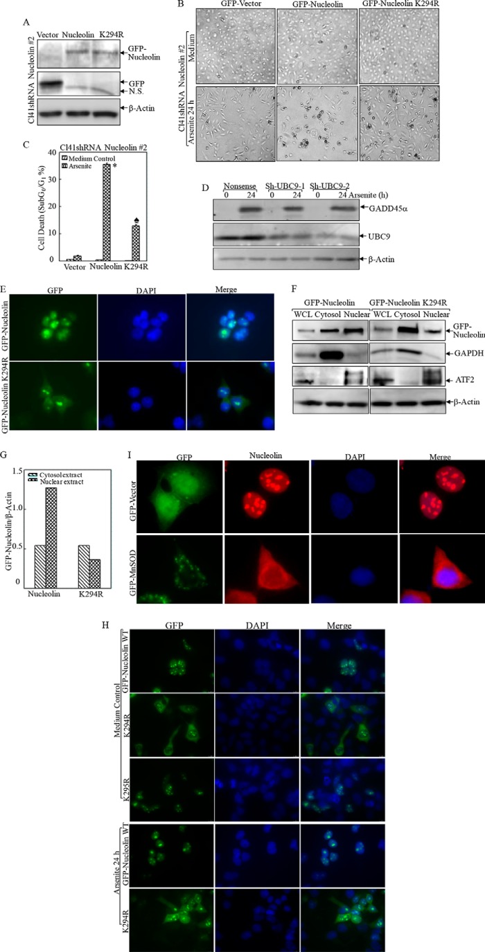
Nucleolin SUMOylation was essential for its nuclear localization and proapoptotic effect following arsenite treatment. A, wild type and K294R mutant human GFP-tagged nucleolin were overexpressed in Cl41 nucleolin shRNA 2 stable transfectants. B and C, the indicated cells were treated with arsenite, and the morphological images were captured under an inverted microscope (B). Cell death was quantified by flow cytometry after propidium iodide staining (C). *, significant increase compared with vector control transfectants (p < 0.05). ♠, significant decrease compared with GFP-nucleolin transfectants (p < 0.05). D, two sets of shRNA UBC9 were stably transfected into MEFs and exposed to arsenite (20 μm) for 24 h. The induction of GADD45α by arsenite exposure was evaluated in the cell extracts obtained from nonsense control and shRNA UBC9 transfectants by Western blotting. E–H, the subcellular distribution of WT or mutant nucleolin was captured by fluorescent microscopy without (E) or with arsenite treatment (H) or extraction of cytosol and nuclear lysis (F). The relative amount of cytosol and nuclear nucleolin protein was quantified by ImageQuant version 5.2 (G). The data shown are representative of three independent experiments. I, the subcellular distribution of nucleolin in the transfectants of GFP-vector and GFP-Mn-SOD was captured by fluorescent microscopy. WCL, whole cell lysis. Error bars, S.D.

