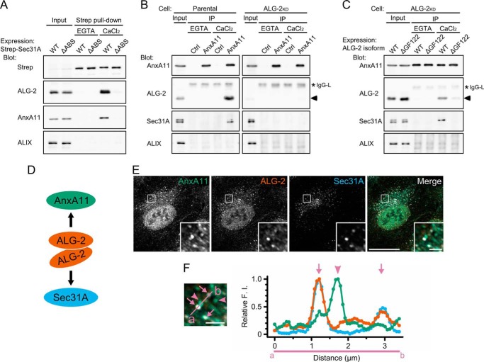
FIGURE 3.
ALG-2 calcium-dependently couples AnxA11 with Sec31A. A, immunoblots of proteins in the Strep pulldown products from HEK293 cells expressing Strep-tagged Sec31A (WT) or its deletion mutant lacking the ALG-2-binding site (amino acids 839–851) (ΔABS). HEK293 cells transfected with pSTREP-Sec31A or pSTREP-Sec31AΔABS were lysed, and cleared lysate (Input) was incubated with Strep-Tactin beads in the presence of 5 mm EGTA or 100 μm CaCl2. After washing, pulldown products (Strep pulldown) were analyzed by Western blotting using indicated antibodies. The relative amount of cleared cell lysate proteins (Input) used for analysis of Strep pulldown products was 20% for Strep, 5% for ALG-2, and 0.5% for AnxA11 and ALIX. Representative data obtained from three independent experiments is shown. B and C, immunoblots of proteins in the immunoprecipitates (IP) with antibodies against AnxA11 from the cleared cell lysates of HEK293 cells (Parental) (B, left panels), ALG-2 knockdown HEK293 cells stably expressing short hairpin-type RNA target for ALG-2 (ALG-2KD) (B, right panels), or ALG-2KD cells transiently expressing either the major or the minor isoform of ALG-2 (WT or ΔGF122, respectively) (C). Cells were lysed, and cleared cell lysate (Input) was incubated first with polyclonal antibody against caspase-1 p20 (negative control, Ctrl) or polyclonal antibody against AnxA11 and then with magnetic beads carrying protein G in the presence of 5 mm EGTA or 100 μm CaCl2. After washing, immunoprecipitates (IP) were analyzed by Western blotting using indicated antibodies. The relative amount of cleared cell lysate proteins (input) used for analysis of immunoprecipitation products was 10% for AnxA11, 3% for ALG-2, and 0.5% for Sec31A and ALIX. Representative data obtained from three (B, left panel, and C) and two (B, right panel) independent experiments are shown. Arrowheads and asterisks indicate ALG-2 and light chains of IgG, respectively. D, schematic representation of ALG-2-mediated physical association of AnxA11 with Sec31A. ALG-2WT forms dimer and couples AnxA11 with Sec31A in a calcium-dependent manner. E and F, confocal micrographs of HT1080 cells stained with antibodies against AnxA11, ALG-2, and Sec31A. Cells were fixed with 4% paraformaldehyde in phosphate buffer on ice for 1 h, permeabilized with 0.1% Triton X-100, and then triple-stained with indicated antibodies. The right panel in E shows merged images with the pseudocolors as follows: green (AnxA11), orange (ALG-2), and light blue (Sec31A). The lower right insets are magnified view of the white squares. Scale bars, 20 μm and 2 μm (inset). Left panel in F is magnified image of inset from right panel in E. Arrows and arrowheads indicate AnxA11-positive puncta overlapping and nonoverlapping with ALG-2/Sec31A-positive structures, respectively. Scale bar, 2 μm. Fluorescence intensities along a line scan indicated in the left panel (from a to b) crossing Sec31A/ALG-2-positive puncta were measured by ImageJ, and relative fluorescence intensities are presented by green (AnxA11), orange (ALG-2), and light blue (Sec31A) in a graph (F, right panel), where the maximum value is 1.0.

