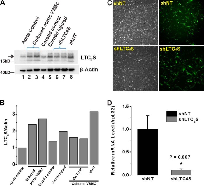
FIGURE 1.
A, LTC4S expression levels are higher in synthetic VSMCs (cultured proliferative aortic VSMCs; lane 2 and 3) compared with acutely isolated contractile/quiescent VSMCs (from medial aorta; lane 1). The LTC4S band is indicated by the arrow. Acutely isolated medial VSMCs from a healthy carotid artery (lane 4) and VSMCs from balloon-injured carotid artery (lane 5; 2 weeks postinjury) were assayed for LTC4S expression; LTC4S protein is up-regulated at 2 weeks after injury. Synthetic cultured aortic VSMCs infected with lentiviral particles encoding either shNT (lane 8) or shLTC4S (lanes 6 and 7) were lysed 7 days postinfection, and LTC4S expression was determined. B, densitometry of LTC4S and β-actin bands from experiment shown in A was performed using NIH ImageJ, and ratios of LTC4S/β-actin are represented. C, cultured synthetic VSMCs were infected with GFP-expressing lentiviral particles encoding either shLTC4S or shNT. Bright field images show total cells, and images of green fluorescence show successfully infected cells. The infection efficiency is >90%. D, VSMCs were infected by either shNT- or shLTC4S-encoding lentiviruses, and RT-PCR analysis was performed using LTC4S-specific primers and normalized to the 60 S ribosomal protein L32 (rpL32) transcript used as an internal control. Data represent the mean ± S.E. (error bars) from three independent infections. *, p < 0.05.

