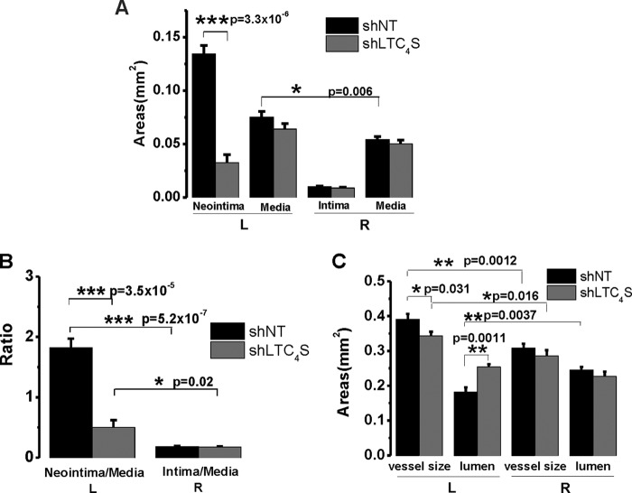
FIGURE 5.
A, statistical analysis of the cross-sectional areas (n = 6 rats per condition). Error bars represent S.E. In injured vessels, the average neointimal area of the shLTC4S group is significantly smaller than that of the shNT control group. The area size of the former is only one-fourth of the size of the latter (0.032 versus 0.134 mm2). The media size was slightly but significantly increased after injury compared with the intact artery (0.075 versus 0.064 mm2). When treated with shLTC4S, the injury-induced increase in media size was slightly attenuated, although this is not statistically significant. B, neointima size was normalized to media size to rule out variations in vessel size among different rats. For both groups, the neointima/media ratio is significantly higher compared with their respective right carotid controls. The neointima/media ratio of shLTC4S group is dramatically lower than that of the shNT group (0.50 versus 1.83). C, the left carotid vessel size was significantly enlarged after balloon injury compared with the intact right carotid artery from the same animal. For the shNT group, the injured artery size is 0.39 mm2, whereas the intact right control artery size is 0.31 mm2. For the shLTC4S group, the injured artery size is 0.34 mm2, whereas the intact right control artery size is 0.29 mm2. The intact artery sizes between these two groups were not significantly different, whereas the injured artery size of the shLTC4S group was significantly lower than that of the shNT group. The lumen was significantly narrowed after injury (0.18 versus 0.24 mm2). When treated with shLTC4S, the vessel lumen size was essentially normalized (0.25 versus 0.23 mm2). *, p < 0.05; **, p < 0.01; ***, p < 0.001.

