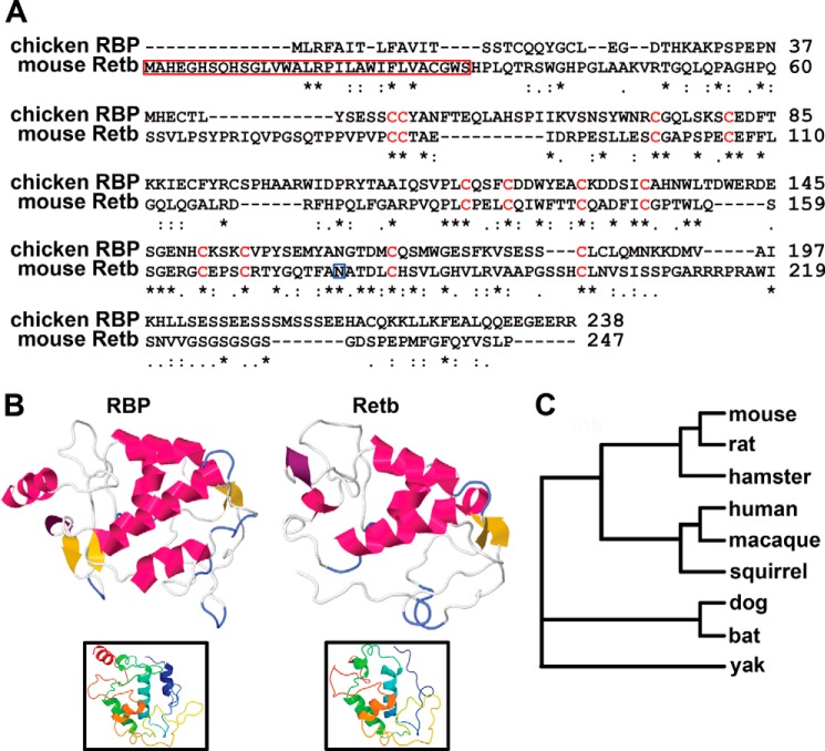
FIGURE 1.
Retb has significant sequence homology to riboflavin-binding protein. A, Clustal Omega alignment of chicken RBP and mouse Retb showing conserved residues indicated by (*), strongly similar by (:), and weakly similar by (.). Conserved cysteines are shown in red. Red box indicates potential cleaved signal sequence, and blue box points to potential N-linked glycosylation site. B, Phyre2 server tertiary structure prediction of RBP and Retb showing helices in pink, β-sheets in yellow, and turns in purple. Insets represent the N-to-C tertiary structure as a rainbow (blue to red). C, STRAP phylogenic tree of the Retb sequence is displayed showing similarity across mammalian species.

