FIGURE 3.
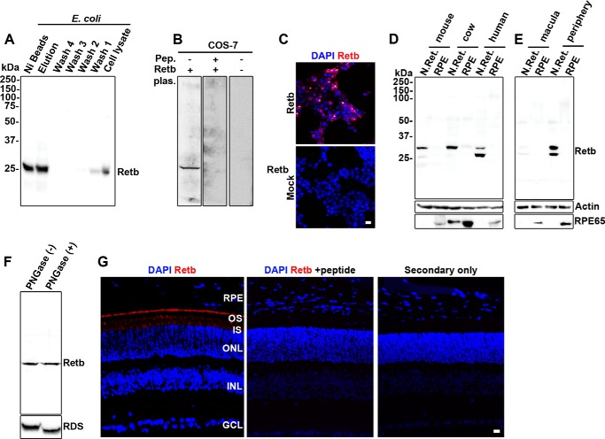
Retb protein is found in the periphery of the neural retina but not the macula or RPE. A–C, specificity of Retb polyclonal antibody was assessed against recombinantly expressed Retb. A, immunoblots of His-tagged full-length Retb eluted from nickel-nitrilotriacetic acid beads using imidazole exhibited a single band of predicted size. B and C, COS-7 cells were transiently transfected with Retb plasmid (or Mock). B, immunoblot showing that Retb antibody signal was completely competed out using 50 μg of the peptide (Pep.) used to generate the antibody. Plasm, plasmid. C, immunocytochemistry showing Retb labeling (red) in COS-7 cells transfected with Retb at ×10, although mock-transfected cells showed no signal (bottom panel) indicating specificity of the antibody to the expressed protein. D, immunoblot of mouse, bovine, and human neural retina (N.Ret.) and RPE (PECS for mice and RPE for human and bovine) protein extracts probed with anti-Retb antibody. A single band of ∼30 kDa is shown in all samples, and a lower band at varying intensities is shown in human and mouse retinal extracts. E, immunoblot of human macula and peripheral neural retina and the corresponding RPE blotted with the anti-Retb antibody. Retb is preferentially expressed in the peripheral human retina than in the macula. F, immunoblot of PNGase F-treated and -untreated P30 neural retinal extracts probed with anti-Retb or anti-Rds antibodies. G, representative IF using the anti-Retb antibody in red (left panel), anti-Retb antibody competed out by preincubation with peptide (middle panel), and secondary antibody only as control (right panel). Nuclei were counterstained with DAPI (blue). OS, outer segment; IS, inner segment; ONL, outer nuclear layer; INL, inner nuclear layer; GCL, ganglion cell layer. Scale bars, 10 μm.

