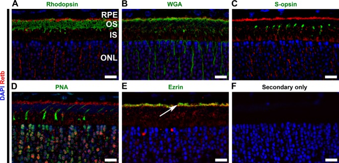
FIGURE 4.
Retb is localized at the interface between the OSs and RPE microvilli. Representative single confocal images of P30 retinal cross-sections were immunolabeled for Retb (red) and different markers (green) for photoreceptor, RPE, and IPM. Nuclei are counterstained with DAPI. A, IF images co-labeled with anti-Retb (red) and anti-rhodopsin (green). Retb is primarily concentrated at the tips of the rod OSs and is apical to rhodopsin. B, IF images co-labeled with Retb (red) and WGA (green). Retb co-localized with WGA. C, IF images co-labeled with Retb (red) and S-opsin (green). No co-localization was seen between Retb and S-opsin. D, IF images co-labeled with Retb (red) and PNA (green). No co-localization was seen between Retb and PNA. E, IF images co-labeled with Retb (red) and ezrin (green). Retb co-localized with ezrin and located basal to the RPE microvilli (indicated by an arrow). F, secondary antibody alone as a control. Nuclei are counterstained with DAPI (blue). Scale bars, 10 μm. The distribution of Retb around the inner segments seems nonuniform, which may be partly due to the fact that those images represent single planes from a deconvolved image stack and that Retb may be localized in individual domains/clusters within the extracellular space. OS, outer segment; IS, inner segment; ONL, outer nuclear layer.

