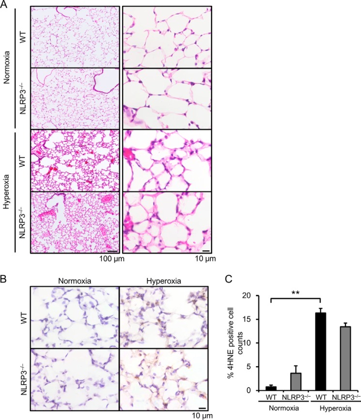
FIGURE 2.
NLRP3−/− mice exhibit similar acute lung injury and generation of reactive oxygen species. Lung samples were obtained from WT and NLRP3−/− mice exposed to normoxia or hyperoxia for 72 h. A, the lung sections were stained with H&E. Representative images of H&E staining are shown (n = 3–4 for each). B, reactive oxygen species generation was assessed by immunohistochemistry with an anti-4-HNE antibody. C, quantitative analysis (n = 3–4 for each). Data are expressed as the means ± S.E. **, p < 0.01.

