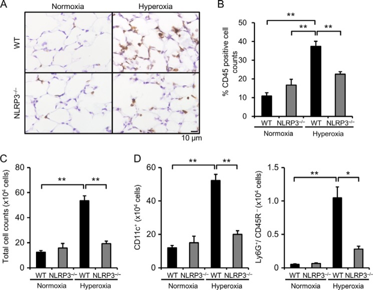
FIGURE 3.
NLRP3−/− mice exhibit less inflammatory cell infiltration. Lung samples were obtained from WT and NLRP3−/− mice exposed to normoxia or hyperoxia for 72 h. A, lung sections were immunohistochemically stained with antibody against CD45. B, quantitative analysis (n = 3–4). C, the total cell count in BALF was determined. D, the number of alveolar macrophages (CD11c+) and neutrophils (Ly6G+/CD45R−) in BALF was analyzed by flow cytometry (n = 3–4 for each in normoxia, and n = 5–6 for each in hyperoxia). Data are expressed as the means ± S.E. *, p < 0.05; **, p < 0.01.

