FIGURE 8.
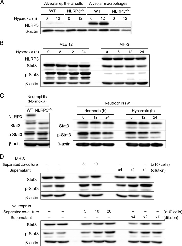
Macrophages and neutrophils increase Stat3 activation in alveolar epithelial cells. A–C, primary alveolar epithelial cells, macrophages, and neutrophils were isolated from WT and NLRP3−/− mice. Alveolar epithelial cells, macrophages, neutrophils, MLE 12 murine lung epithelial cells, and MH-S murine alveolar macrophage cells were exposed to normoxia or hyperoxia for the indicated times. A, Western blot analysis of NLRP3 expression in alveolar epithelial cells and macrophages (n = 3). B and C, Western blot analysis of NLRP3 expression and Stat3 expression and activation using antibodies against Stat3 and phospho-Stat3 (p-Stat3). D, MLE 12 cells were separately co-cultured with MH-S cells (5–10 × 105) or neutrophils (5–20 × 105) for 8 h. The supernatants from MH-S cells and neutrophils (diluted ×4 and ×2) were added to MLE 12 cells and incubated for 8 h. Shown are the results from Western blot analysis of Stat3 expression and activation in MLE 12 cells using antibodies against Stat3 and phospho-Stat3.

