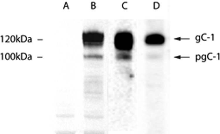
FIGURE 6.

Crude cell lysate-derived gC-1 and gC-1 isolated by immunoaffinity chromatography visualized by immunoblot. Lane A, uninfected cell lysate. Lane B, HSV-1-infected HEL cell lysate. Lane C, gC isolated from HSV-1 infected HEL cells. Lane D, gC isolated from released HSV-1 viral particles. Positions of molecular weight markers and fully processed gC-1, containing complex type N-linked glycans and a variety of O-linked glycans, and precursor gC-1 (pgC), containing high mannose N-linked glycans and neglible O-linked glycans. The differences between gC-1 and pgC-1 are reviewed elsewhere (9, 14).
