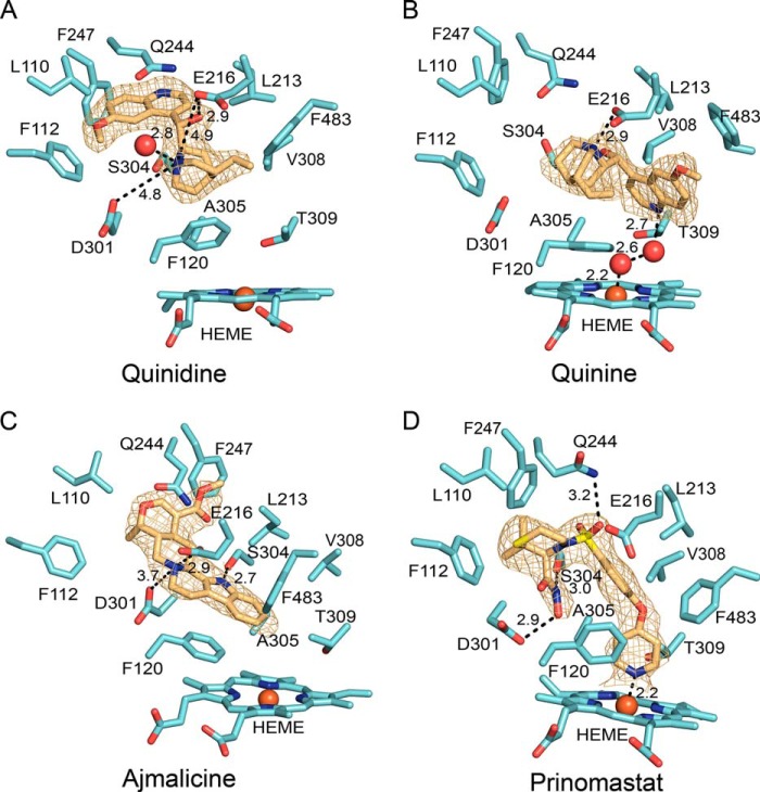
FIGURE 4.
Binding of quinidine (A), quinine (B), ajmalicine (C), and prinomastat (D) in the active site of P450 2D6 after competitive displacement of thioridazine in crystals of the co-crystallized P450 2D6 ternary thioridazine complex. The gold mesh depicts 2m|Fo| − D|Fc| ligand omit maps contoured at 1 σ around the ligand. Distances between protonated nitrogens and Asp-301 and/or Glu-216 as well as hydrogen bonds and the coordinate covalent bond of the pyridine moiety of prinomastat or water to the heme iron are depicted by dashed lines with the distances shown in Å. P450 carbons are colored cyan, and ligand carbons are colored amber. Other atoms are colored as described in the legend to Fig. 2.

