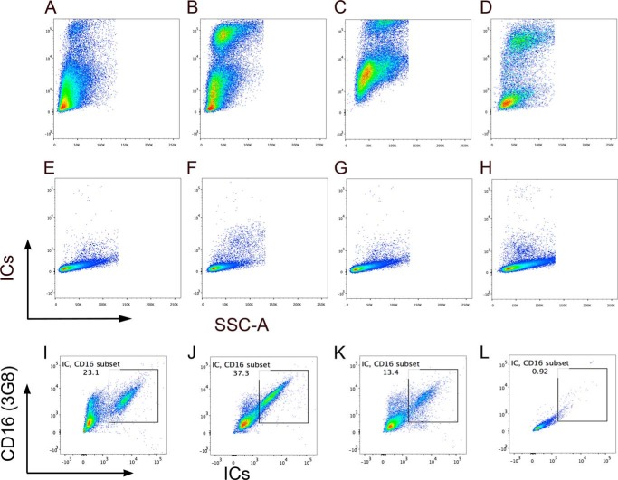
FIGURE 3.
Binding of IC-Alexa-Fluor 488. Activated CD4+ T-cells Jurkat (A), P116 (B), and monocytic cells THP-1 (C) as well as Daudi B cells (D) demonstrate binding to labeled ICs. Unstained Jurkat (E), P116 (F), THP-1 (G), and Daudi (H). Dual binding of ICs and anti-CD16 (3G8). THP-1 (I), activated P116 (J), and activated Jurkat (K) and unstained cells (L). Cells stained with anti-CD16 also show ICs staining. Data representative of two independent experiments.

