
FIGURE 3.
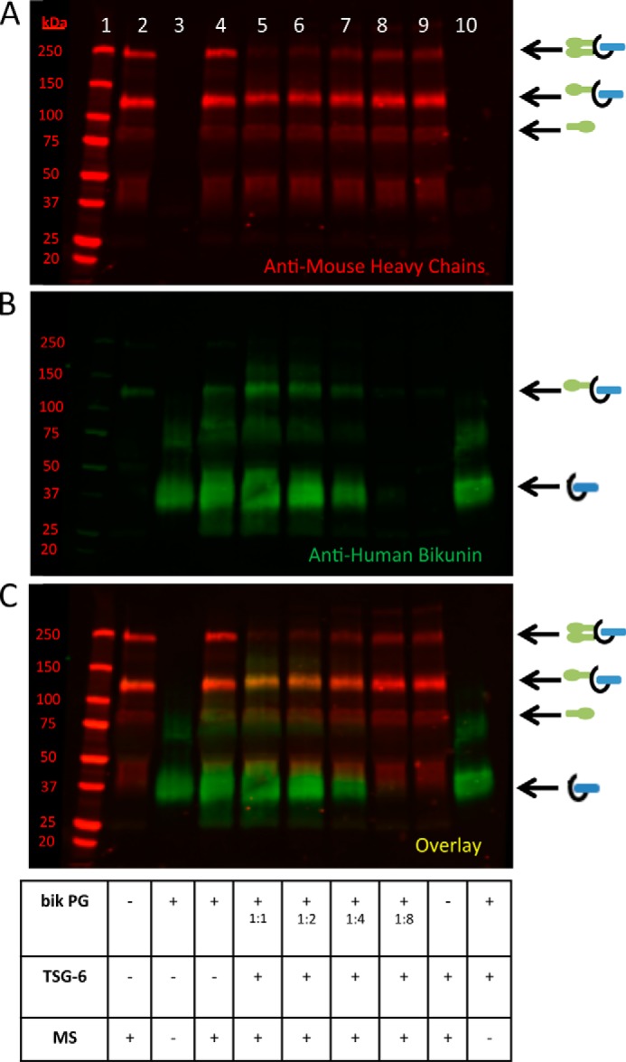
A portion of the bikunin proteoglycan is unable to accept heavy chains. Shown is a Western blot of samples containing mouse serum incubated at 37 °C for 4 h with recombinant TSG-6 and the purified bikunin PG, as listed in the table. A–C represent the same blot, which was probed with an anti-human bikunin antibody (green) and anti-mouse antibodies for HCs 1 and 2 (red). A portrays the red channel only, B portrays the green channel only, and C portrays the overlay. Lane 1 portrays the Mr standards. The sample in lane 2 contains mouse serum alone. A 1:2 serial dilution of the bikunin PG is portrayed in lanes 5–8. Recombinant human TSG-6 was added to the samples in lanes 5–10. The sample in lane 3 contains the bikunin PG only, and the sample lane 10 contains both the PG and TSG-6. MS, mouse serum.