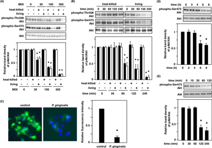
FIGURE 1.
P. gingivalis dephosphorylates Akt at Thr-308 and Ser-473. A and B, Ca9-22 cells were infected with heat-killed or live P. gingivalis. A, MOI dependence of Akt dephosphorylation for 2 h. B, infection time dependence of Akt dephosphorylation at an MOI of 100. After incubation, the cells were lysed and analyzed with SDS-PAGE and Western blotting probed with the indicated antibodies. The graphs below indicate the ratio of phospho-Akt at Thr-308 (solid bar) and Ser-473 (open bar) to total Akt by Western blotting. C, Ca9-22 cells were infected with or without P. gingivalis at an MOI of 100 for 2 h. The infected cells were fixed with 4% paraformaldehyde in PBS, and immunofluorescence was evaluated as described under “Experimental Procedures.” The graph indicates the relative fluorescence intensity for the phospho-Akt positive cells compared with control cells. D and E, Ca9-22 cells in 24-well plates were incubated with P. gingivalis at an MOI of 300 for up to 8 h through a cell culture insert system (D), and Ca9-22 cells were incubated for 120 min with P. gingivalis culture supernatant prepared as described under “Experimental Procedures” (E). After incubation, the cells were lysed and analyzed with SDS-PAGE and Western blotting probed with the indicated antibodies. The graphs indicate the ratio of Ser(P)-473 Akt/total Akt on Western blotting. These results were independently demonstrated on three separate occasions. Statistical significance: *, p < 0.05.

