FIGURE 2.
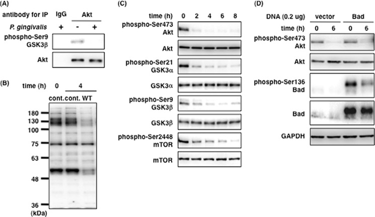
P. gingivalis induces Akt inactivation and the dephosphorylation of the downstream proteins in the PI3K/Akt signaling pathway. A, Ca9-22 cells were infected with or without P. gingivalis at an MOI of 100 for 4 h. Cell lysates were incubated with an anti-Akt monoclonal antibody immobilized with Sepharose beads for 4 h. Akt kinase assay was performed as described under “Experimental Procedures.” B, Ca9-22 cells were infected with or without P. gingivalis at an MOI of 100 for 4 h and prepared for whole cell lysates. The lysates were analyzed with SDS-PAGE and Western blotting using anti-Ser(P)/Thr(P) Akt substrate antibody. C and D, Ca9-22 cells were infected with P. gingivalis at an MOI of 100 for up to 8 h (C), and Ca9-22 cells were transfected with 0.2 μg of pEF4C-His-Bad. After transfection, Ca9-22 cells expressing His-Bad were infected with P. gingivalis at an MOI of 100 for 6 h (D). The infected cells were lysed and analyzed with SDS-PAGE and Western blotting probed with the indicated antibodies, respectively (C and D). These results were independently demonstrated three times. cont., control; IP, immunoprecipitation.

