
FIGURE 3.
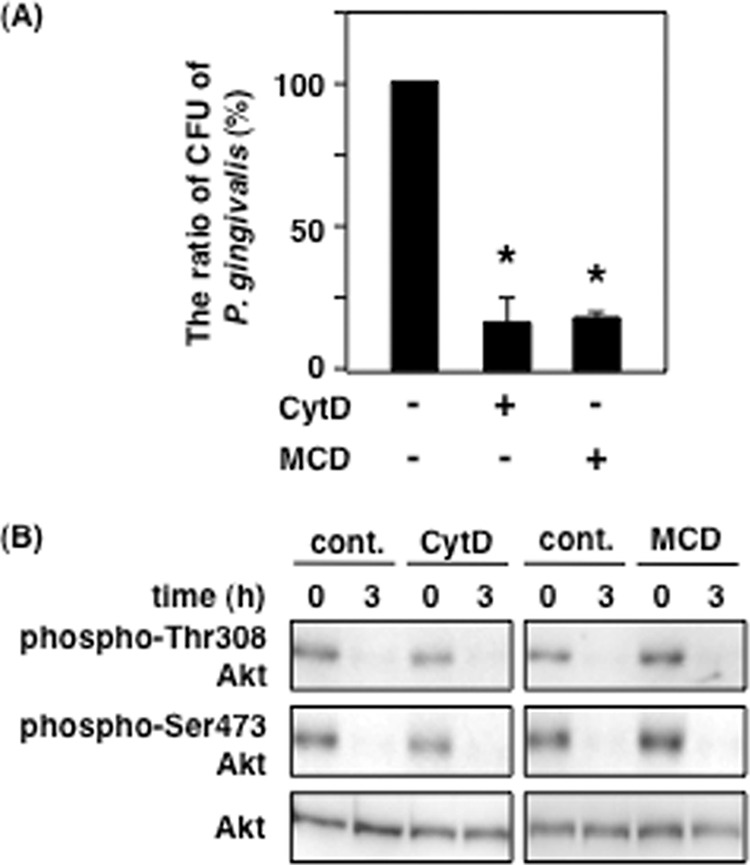
Effect of P. gingivalis invasion on P. gingivalis-induced Akt inactivation. Ca9-22 cells were treated with or without 10 μm CytD or 1 mm MCD for 1 h. A, the treated cells were infected with P. gingivalis at an MOI of 100 for 2 h. P. gingivalis invasion assay was performed using the antibiotics treatment as described under “Experimental Procedures.” B, the treated cells were infected with P. gingivalis at an MOI of 100 for 3 h and then lysed. The lysates were analyzed with SDS-PAGE and Western blotting using the antibodies for total Akt and phosphorylated Akt at Thr-308 and Ser-473. These results were independently demonstrated three times. Statistical significance: *, p < 0.05. cont., control.