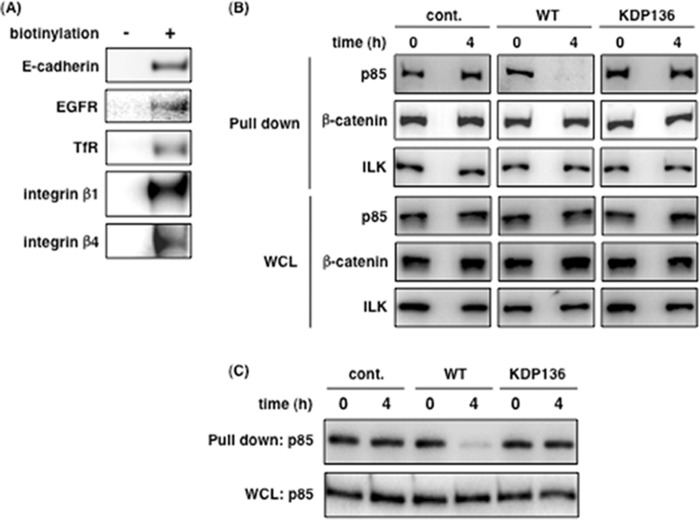
FIGURE 5.
Interaction between PI3K p85α and membrane proteins is disrupted by gingipains. A, all membrane proteins in Ca9-22 cells were labeled with biotin for 30 min, and the cells were lysed for a pulldown assay as described under “Experimental Procedures.” The pulldown samples were then analyzed by SDS-PAGE and Western blotting using the antibodies for the respective membrane proteins. B and C, untreated Ca9-22 cells (B) and 10 μm CytD-treated Ca9-22 cells (C) were infected with or without P. gingivalis WT or KDP136 at an MOI of 100 for 4 h. After infection, the cells were labeled for 30 min, lysed, and run in a pulldown assay. The precipitants were analyzed by SDS-PAGE and Western blotting using the indicated antibodies. These results were independently demonstrated three times. cont., control; WCL, whole cell lysate; ILK, integrin-linked kinase.

